Radiology Imaging Services
 We offer comprehensive imaging options, including:
CT Scanning This is computed axial imaging of all parts of the body utilizing a CT scanner. Some of the applications of CT scanning are brain/head, chest, heart, abdominal, bone-density scans and CT angiography using multi detector technology. UTMC offers imaging from the Toshiba Aquilion ONE ViSION.
CT Angiograms Help diagnose blood vessel blockages, identify blood-rich tumors or help locate where a blood vessel may be bleeding. Blocked blood vessels can be treated by cardiologists or vascular surgeons as soon as the diagnosis is made.
Diagnostic Services Include general x-ray work such as chest and bone x-rays, as well as a full spectrum of upper GI, lower GI studies.
Interventional Neuro-Radiology We can perform Intracranial thrombolysis for stroke, and intravascular treatment and embolization of intracranial Berry Aneurysm, using unique advanced imaging equipment. This allows the interventionalist to work from within blood vessles inside the brain to treat intracranial aneurysms, remove obstructions in acute stroke, and repair vascular malformations. We work closely with neurosurgeons to treat patients prior to surgery to prevent intraoperative complications.
We have a comprehensive spine care team to care for patients using the latest techniques and treatments. There are several interventions for back pain resulting from compression fractures and nerve damage. Epidural injections and nerve blocks are some of the most effective treatments for relieving back pain and treating pinched or irritated nerves. Kyphoplasty and verterbroplasty are performed to help eliminate pain from compression fractures by treating the collapsed vertebral bodies.
Interventional Radiology UTMC Interventional Radiologists use minimally invasive techniques in conjunction with imaging, such as CT Scan, MRI Scan and Ultrasound to guide instruments through the body and directly to the area of interest. The benefits of utilizing a minimally invasive procedure, instead of surgery are less risk, less pain and reduced recovery time. Most often they are done on an outpatient basis, allowing you to return home the same day and require little to no anesthesia. UTMC performs many interventional procedures such as biopsies, drain abscesses, fine needle aspirations, thoracentesis, and paracentesis.
Kyphoplasty and Vertebroplasty Two procedures recommended for compression fractures. Both procedures use bone cement to improve stability and treat pain by raising collapsed bones or filling hairline cracks in bone.
Magnetic Resonance Imaging Utilizes a high strength magnetic field and radio-frequency waves to assess central nervous system disorders, musculoskeletal disease and sports medicine injuries, heart and great vessel disorders, abdominal/pelvic masses and vascular disease, among others. The UT Medical Center is home to both a 3.0T MRI unit and a 1.5T MRI unit.
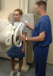
Mammograms /Breast Exams These exams are performed utilizing Breast tomosynthesis, which is the latest in mammography technology. Breast tomosynthesis, also called 3-D mammography can produce three-dimensional images of the breast, which leads to better cancer screening and detection. Using this procedure, the breast is viewed as many thin slices, which can be combined into a 3-D picture. It can be used to investigate the cause of breast problems, such as breast mass, pain and nipple discharge.
The benefits of 3-D mammography include:
- Better visualization leading to earlier detection of abnormalities in the breast
- Clearer images provide a greater level of detail in examining your breast
- Fewer unnecessary biopsies
Nuclear Medicine Images the human body by injecting radiopharmaceuticals, (isotopes) typically into the patient’s vein and creating an image of the path that they make. These images are used to determine function or flow of organs/systems. Typical exams are bone, lung, and liver scans. Molecular medicine routinely utilizes two different imaging techniques (SPECT and PET). When combined, the images show details of an organ’s anatomy as well as how it’s functioning. These studies help surgeons treat cancer and sometimes prevent the need for invasive surgery. Exposure to radiation is low, quick, and carefully controlled.
Ultrasound Images the internal structures of the human body by sending high-frequency sound waves through the skin which sends back echoes, producing an image. Exam times vary from 15 minutes to an hour. Ultrasound has become the premier technology for fetal (pregnancy) scans, gallbladders, kidney scans, peripheral vessels, and breast (cysts). Doppler ultrasound allows the radiologist to evaluate the function of the patient’s carotid arteries and risk of developing stroke or cerebral vascular disease.